

The title page of the 1523 Bologna edition. Photograph: National Library of Medicine.
Jacopo Berengario da Carpi (also Jacobus Berengarius Carpensis, Jacopo Barigazzi, or simply Carpus) was born in Carpi, near Modena, around 1460, the son of a surgeon. As a young man he studied under the famous printer and editor Aldus Manutius. He trained in medicine at Bologna, taught surgery at Pavia, and from 1502 to 1527 was on the faculty at Bologna. He also lived at times in Ferrara and Rome, where he was among the first to treat syphilis with mercury.
Anatomy was his passion. He boasted of having dissected several hundred bodies, and his enemies went so far as to accuse him of dissecting living people. His skill was known across Italy; he was invited to become the Pope's physician and declined. He died in Ferrara around 1530, a wealthy man, and left his fortune to Duke Alfonso I of Ferrara, husband of Lucrezia Borgia.
Beyond Mondino
Anatomy in Berengario's day still meant Mondino dei Luzzi (died 1326), whose text relied mostly on Arab physicians and a few dissections, and was never illustrated. Berengario revered it, but he was an eager, tireless observer himself, and he is credited with the first anatomical illustrations made from nature.
His first illustrated book, a commentary on Mondino's Anatomia, came out in Bologna in 1521. In 1522 and 1523 he published the Isagogae breves ("short introductions"), a compendium meant to replace Mondino's book, which it far outshone. The 1523 edition shown here adds four illustrations of the heart and two of the brain, and varies some of the muscle woodcuts. Some think Berengario had no hand in those new pictures, but no one can be sure; others have suggested that the woodcuts in both his books were made by the artist Hugo da Carpi (1455–1523).
The plates
The muscle figures show the body opening itself up: a man lifts his own skin to show the muscles of his belly, then peels it wider to show the obliques and arms.
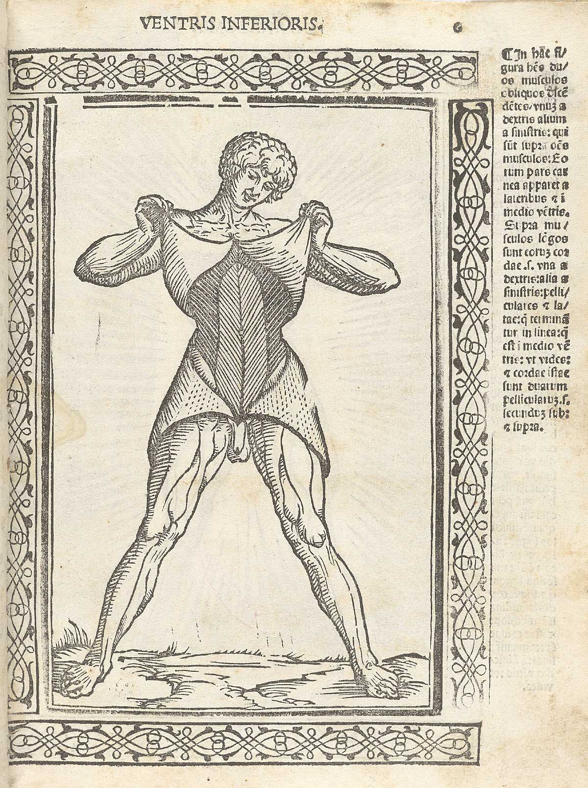
A figure lifts his skin to show the abdominal muscles. Photograph: National Library of Medicine.

Pulling the skin aside. Photograph: National Library of Medicine.
Other plates show the organs: a seated woman with her abdomen opened, in a room with a window onto a city; the uterus, inside and out; the heart in cross-section, with its chambers and great vessels; and the brain from above and below.

The female reproductive organs, shown in a domestic interior. Photograph: National Library of Medicine.

The heart in cross-section, one of the 1523 edition’s additions. Photograph: National Library of Medicine.

The brain from above and below. Photograph: National Library of Medicine.
The book ends with the frame: the numbered vertebrae of the spine, the veins and arteries of the arm, the muscles of standing figures, and skeletons posed front and back.

The spine, vertebra by vertebra. Photograph: National Library of Medicine.

A skeleton, from the front. Photograph: National Library of Medicine.
Sources
- National Library of Medicine, Historical Anatomies on the Web: Jacopo Berengario da Carpi, Isagogae breues, perlucidae ac uberrimae, in anatomiam humani corporis (Bologna: Benedictus Hector, 1523). Further reading there: L. Choulant, History and Bibliography of Anatomic Illustration; N. G. De Santo, A. Touwaide and others, "Berengario da Carpi," American Journal of Nephrology; L. Merlini, P. Tomba and A. Vigano, "Berengario da Carpi, a pioneer in anatomy, rediscovered by Vittorio Putti," Neuromuscular Disorders; Morton's Medical Bibliography.
Licence: CC0 1.0 (public domain) · Adapted from www.nlm.nih.gov
1
0
0
0

Comments


