Viu algo para melhorar? Proponha uma alteração.
Esta página é pública.
Introduction
Cell adhesion is fundamental in the formation of tissues due to the attraction between individual cells. These adhesions are due to the internal cytoskeleton, which determines the overall structure of the cell. The cell can be in two states - stable adhesion interactions and dynamic adhesive interactions. The stable adhesion interactions are typically made up of cell adhesion receptors (which usually are the glycoprotein that binds the ECM between cells), ECM (extracellular matrix, which are large proteins that interact with other cellular receptors on the site) proteins, and cytoplasmic plaque proteins.
Formation of Tissue
One of the most significant cell adhesions that is required to form a stable tissue is called the Cadherins adhesion molecules, which are transmembrane receptors that depends on Ca2+ to recognize other cells during growth. One of the most studied cadherin molecules is called E-cadherin, which is known to fight epithelial cancer. Cadherin work by combining with catenins and the actin in the cytoskeleton. Catenins are cytoplasmic plaque proteins, which incorporates both a-catenins and b-catenins. a-catenins are responsible for the linkage between the cadherins to the actin in the cytoskeleton. b-catenins are responsible for the interaction between the a-catenin and the cadherin cytoplasmic domain. Based on the observations of b-catenin's interaction with growth factors and transformation, it can be concluded that b-catenin are simply acting as the regulatory component. Cadherins are also responsible for the regions of the cell known as the junctional localization, which are the stronger points of the cell that is capable for adhesion. The EC (extracellular matrix) domains are split into five sub-domains, EC1, EC2, EC3, EC4, and EC5. With the help of x-ray crystallography, the EC1 sub-domain can be used to form a dimer, more specifically called the strand dimer, which are monomers that are parallel to the adhesive binding surfaces and orient outwards.
Desmosomal Junctions
Desmosomal junctions are primarily located in epithelia and cardiac muscles. They link with the intermediate filament cytoskeleton to form a network capable of withholding immense weight.
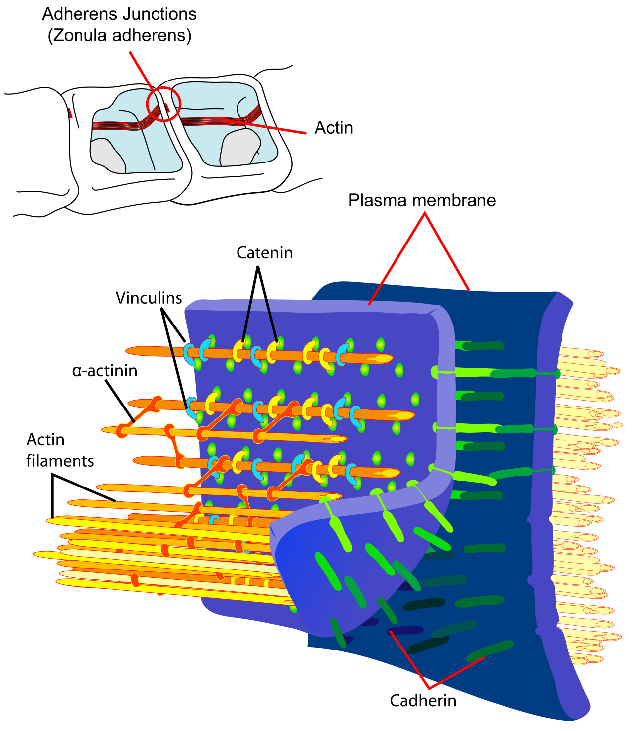
(Image: Mariana Ruiz LadyofHats, Public domain)
References
- Gumbiner, Barry M (1996). "Cell Adhesion: The Molecular Basis of Tissue Architecture and Morphogenesis". Cell. 84 (3): 345–57. doi:10.1016/S0092-8674(00)81279-9.
Where this page came from
This page was imported from Wikibooks. From “Structural Biochemistry” on Wikibooks, by its contributors, under CC BY-SA 4.0. Changed here: set as a page; navigation and edit links left out; each image under its own licence, credited in its caption.
Nobody has written it yet — it is the source material at a new address, which is why search engines are asked to skip it and why no one earns from it. It is up for grabs: take it on, and it is yours to rewrite and to earn from.
Nestas publicaçõesStructural Biochemistry
Licença: CC BY-SA 4.0 · Adaptado de en.wikibooks.org
Comentários